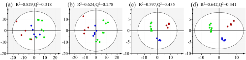
(a) binning results of 1H NMR spectra with normalization to unit weight,
(b) binning results of 1H NMR spectra with normalization to constant sum,
(c) metabolites concentrations obtained by spectral deconvolution and normalization to unit weight,
(d) metabolites concentrations obtained by spectral deconvolution and normalization to constant sum