|
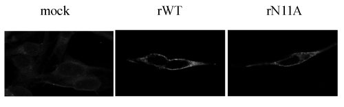
| Figure 3: Immunofluorescence for CM2. HMV-II cells were infected with rWT or rN11A, and incubated for 48 hrs. The cells were stained with anti-CM2 serum and a fluorescein isothiocyanated anti-rabbit IgG, as the primary and secondary antibodies, respectively. The cells were examined by confocal microscopy. |