 |
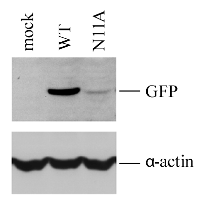
| Figure 4: GFP expression in VLP-infected HMV-II cells. HMV-II cells were infected with WT- or N11A-VLPs, followed by superinfection with AA/50, and then incubated for 48 hrs. The cells were analyzed by immunoblotting using anti-GFP MAb and anti α-actin Ab. |