 |
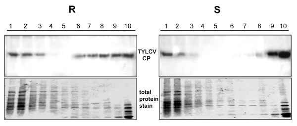
| Figure 1: Distribution of TYLCV CP aggregates following sedimentation on linear 10-50% sucrose gradients. Leaf homogenates were prepared from R and S plants at 28 dpi. Gradients were divided into 10 fractions, 1 (top) to 10 (bottom). Aliquots were subjected to 12% SDS-PAGE. The gels were stained with Coomassie blue (total protein) and western blotted. CP was detected using anti-CP antibody. |