|
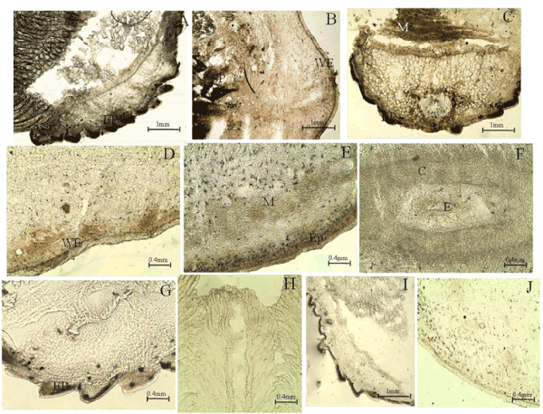
| Figure 1: TUNEL staining for apoptotic analysis in tail regenerates of control H.flaviviridis. (A) Apoptotic activity in the muscle bundles and the keratinized epithelium of the intact tailsegment 48hpa (B) Wound epithelium (WE) stage with apoptosis mainly near the epithelium and nervous tissue with sparse staining in the cell accumulation prior to formation of blastema (BL) (C) Apoptosis in the intact tail segment at the WE stage (D) Localized areas of apoptosis during the BL stage (E) Increased apoptosis in the WE and developing muscle bundles during differentiation (DF) (F) Slight apoptotic activity in the ependyma at DF stage(G) Apoptosis still visible in the keratinized epithelium at later growth stages (H-J) Negativecontrol. |