 |
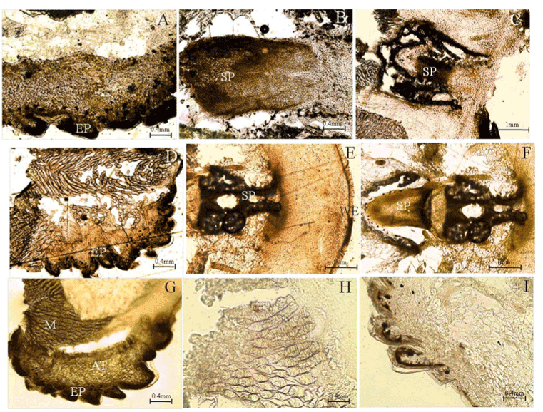
| Figure 2: TUNEL staining for apoptotic analysis in tail regenerates of SU5402 treated H. flaviviridis. (A) Apoptotic activity in the muscle bundles and the connective tissue of the intact tailsegment 48hpa (B) Intense apoptosis staining in the spinal cord 48 hpa(C) Apoptosis after 6dpa (D) Apoptotic staining in the tail 9dpa (E) Increased apoptosis in the WE, blastemal mesenchyme beneath it and spinal cord 11 dpa (F) Intense apoptosis in the nervous tissue (G) Apoptosis in intact tail region 11 dpa (H-I) Negative control AT-adipose tissue, EP-epithelium, M-muscle, SP-spinal cord, WE-wound epithelium. |