|
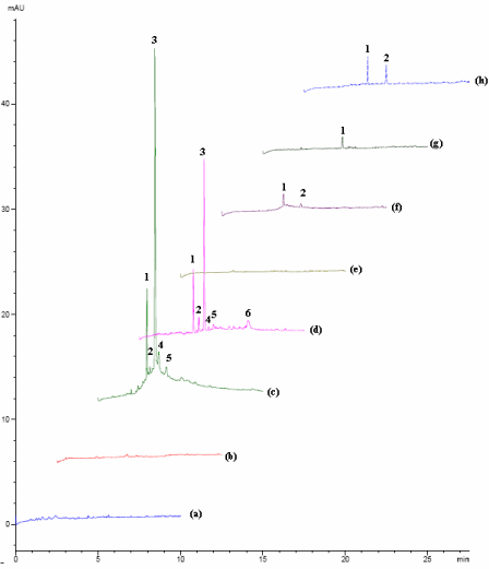
| Figure 4: Electropherograms of force degraded samples under different stress conditions at 257 nm: (a) Standard solution of heparin, (b) Thermal stress at 90°C after 24 hours, (c) Thermal stress at 90°C after 600 hours, 1, 2, 3, 4, 5: degraded forms. (d) Thermal stress at 90°C after 2000 hours, 1, 2, 3, 4, 5, and 6: degraded forms. (e) Acidic hydrolysis (pH=2.00) at 70 ± 1°C for 24 hours, (f) Acidic hydrolysis (pH=2.00) at 70 ± 1°C for 72 hours, 1, 2: degraded forms (g) Basic hydrolysis (pH=12.00) at 70 ± 1°C for 24 hours, 1: degraded form. (h) Basic hydrolysis (pH=12.00) at 70 ± 1°C for 72 hours, 1, 2: degraded form. Despite (b) which was diluted 5 times, all samples were diluted 10 fold before analysis by the optimized method. |