 |
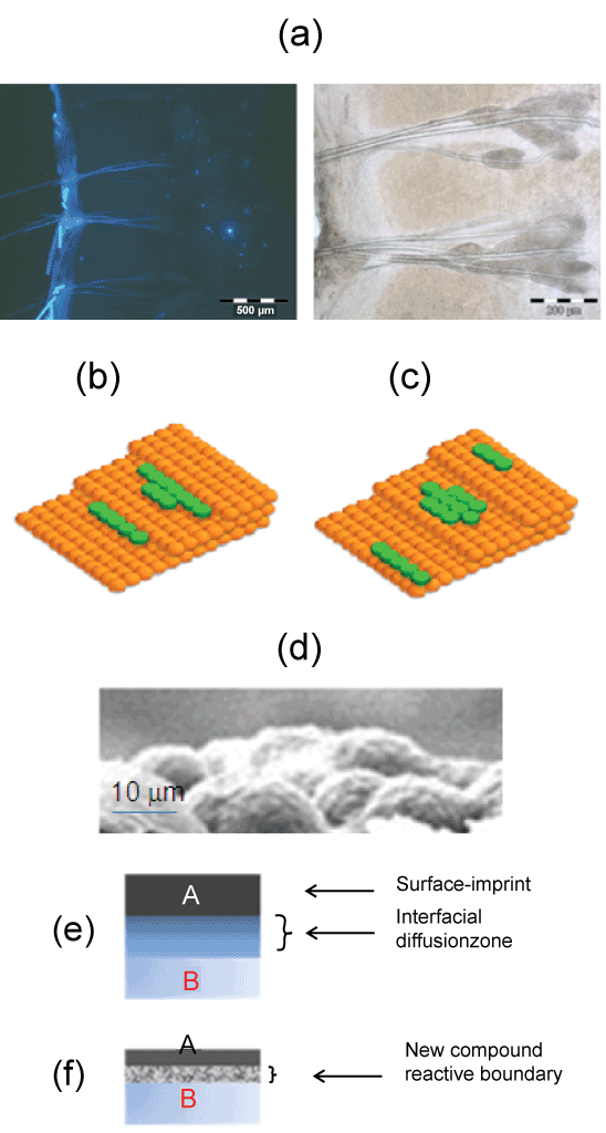
| Figure 3: Top: illustration of topography and scanning electron microscope of microspherical MIP. (a) Left: fluorescent image of for rat skin dorsal represent image of cell receptors and enzyme activity led to fluorescence can be detected by fluorescent microscopy method, right: compared to the bright field image of rat skin [58]. (b) the relative rate of diffusion, accommodation and deposition determine the interfacial interaction and morphology on the barrier kinetic balance of a surface process where facile transport of the delivered substance no interlayer transport occurs and diffusion is rapid before attach of molecules and materials can occur in starking contrast (c) step down diffusion the intermolecular forces between the delivered species is rapid with high interlayer transport. (d) the hierarchically ordered surfaces forming between MIP nanostructures and cast membrane by self-assembly polymerization, showing ordered solid/liquid coexistence. Bottom: Two possible interfaces that can be formed between a membrane consisted of MIP materials (e.g. thin-layer or microparticle) onto the skin: (e) non-abrupt interface and (f) reactive interface [69]. |