|
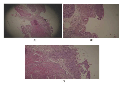
| Figure 13: (A, B and C), shows the micoroscopical features of ulcered mucosa at day 7, the loss of epithelia is clearly evident in A (arrow indicated the remnant of some epithelia), in B the blood vessels, nerve bundles exposed (see the arrow), in C ulceration and poorly definition of the epithelial layer in the control groups (see the arrow) [(H&E stain ×40 for (A) and ×100 for (B and C)]. |