|
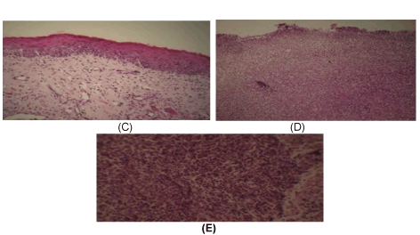
| Figure 20: Microscopical feature of buccal mucosa in the control groups at day 14. The partially healed buccal mucosa is evident in C picture. Extensive necrotic tissue and inflammatory cells in D and E. [H&E stain × 100 for (C and D) and × 400 for (E)]. |