 |
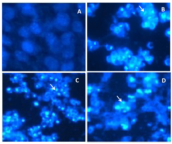
| Figure 4: Induction of apoptotic nuclear morphology of HeLa cells treated with F. solani organic extract. Nuclear morphology was analyzed by fluorescence microscopy upon staining with Hoechst 33342. HeLa cells were cultured in the absence (A) or presence of 2.5 μg/mL (B), 5 μg/mL (C) and 10 μg/mL (D) of F. solani organic extract for 24 h, stained with Hoechst 33342 and analysed as described in Materials and Methods. Arrows indicate the nuclear chromatin condensation foci. |