|
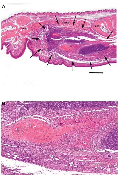
| Figure 2: Photomicrographs of a sagittally sectioned foot of a representative CH3 mouse sacrificed 25 days PI with L. major and treatment with PBS. Panel A) Moderate chronic inflammation is present in the soft tissues (arrows). The footpad epidermis and haired skin are intact. The inflammation has not spread to the bones. H&E, bar = 500 µm. Panel B) The subcutis has sheets of closely packed mixed mononuclear inflammatory cells with darkly stained lymphocytes and plasma cells in the upper half of the photomicrograph and more lightly stained macrophages in the lower half. These surround a central area of dense collagenous connective tissue. H&E, bar = 50 µm. |