|
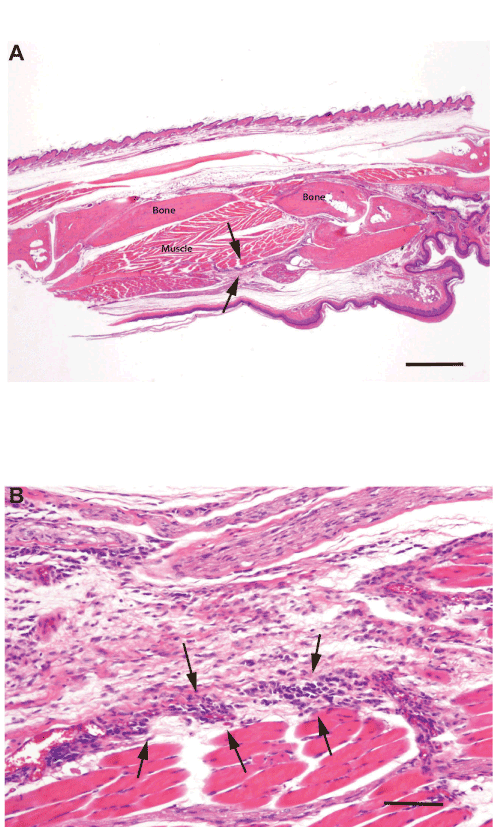
| Figure 3: Photomicrographs of a sagittally sectioned foot of a representative Balb/c mouse sacrificed 25 days following inoculation with L. major and treatment with AMB-ND. Panel A) Note the mild inflammation in subcutaneous tissue (arrows). The footpad epidermis (bottom) is intact and bone and muscle have no lesions. H&E, bar=500 µm. Panel B). The mild focal inflammatory cell infiltrate consists of mixed mononuclear cells (arrows). Normal skeletal muscle lies beneath these cells with normal loose connective above. H&E, bar=500 µm. |