|
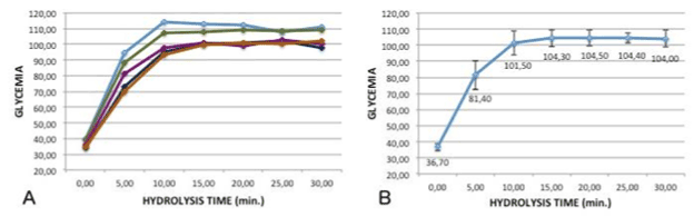
| Figure 1: A: glycemic curves obtained by treating samples of plasma containing fixed concentrations of HES for different times. Each curve represents the analysis of a different sample. B: mean glucose values obtained by treating HES containing blood samples for different periods. A stable and long-lasting plateau was reached after 15 minutes of hydrolysis. Error bars represent 95% confidence intervals. |