|
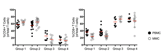
| Figure 1: Impact of IDU on T cell subsets in the peripheral blood and the GI tract. (a) Percentage of CD4+ T cells (left panel) and CD8+ T cells (right panel) from Group 1 (non-IDUs, HIVuninfected, n=13); Group 2 (HIV-1 uninfected IDUs, n=19); Group 3 (HIV-1 infected, viremic, non-IDUs, n=8) and Group 4 (HIV-1 infected, viremic, IDUs n=10). PBMCs are depicted by dark circles and MMC by open grey circles. Red horizontal bars represent the mean per group examined. |