|
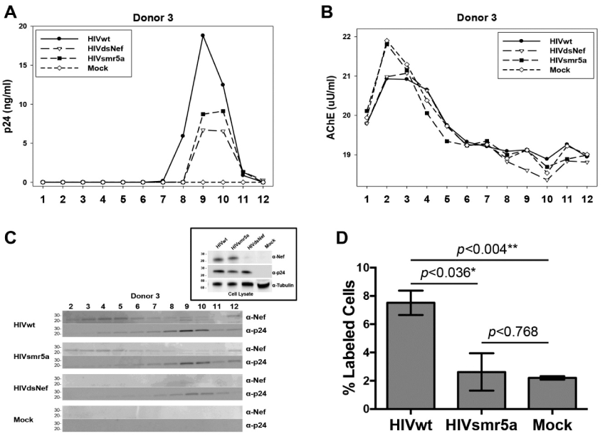
| Figure 5: ExNef is released from HIVNefsmr5a infected cells. (A) 4.0 x 10⁶ HIV seronegative PBMCs were infected with HIVwt, HIVdsNef, or HIVNefsmr5a virus. 12 days post-infection, conditioned cell-free media was harvested and subjected to differential centrifugation at 30,000 × g for 30min and 100,000 × g for 2h. The 100,000 × g pellet was re-suspended in 1 ml of 1X PBS, loaded onto OptiprepTM velocity separation gradient, and subjected to flotation centrifugation at 250,000 × g for 2h. (A-C) Fractions were assayed by p24 and AChE enzymatic assay, a marker for exosomes, and western analysis for Nef and p24 to confirm purification of exosomes from viral particles. While Nef was not detected in the exosome, or the viral fractions of supernatants collected from cells infected with HIVdsNef, Nef was detected in both the exosome, and the viral fractions of supernatants collected from cells infected with HIVwt and HIVNefsmr5a. Plots shown are a representative image from one donor of three independent experiments (D) Exosomes isolated from donor #3 was screened for apoptosis by TUNEL assay. Exosomes were added to target cell cultures at 30 ng/ml as measured by total protein. Compared to exNef from HIVwt infected cells, there was a significant reduction in the number of cells undergoing apoptosis after 48h of exposure to exNef from HIVNefsmr5a infected cells. Error bars represent SEM from the mean. Differences between groups were determined using an unpaired student t-test *p<0.05, **p<0.01. |