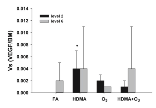
The volume of VEGF positive epithelial cells normalized to surface of basement membrane is represented for all four exposure groups at proximal (level 2) and distal (level 6) airway levels. The HDMA group had significant higher volume of epithelial VEGF expression compared to filtered air at the proximal airway level (*p<0.05).