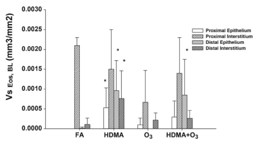
The volume of eosinophils normalized to the surface of the basement membrane in the epithelial compartment and interstitium is represented. The volume of eosinophils was significantly greater in the proximal and distal epithelium as well distal interstitium for the HDMA group and distal epithelium for the HDMA/ Ozone group. (*signifies p<0.05)