|
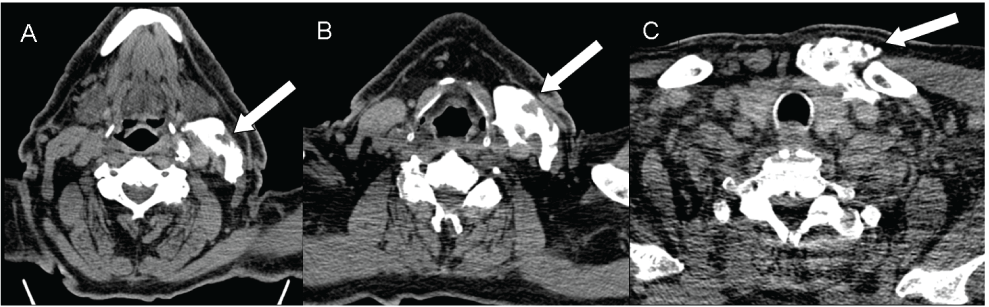
| Figure 1: Axial native CT images of the left sided contrast media/anaesthetic injection at three different levels of the neck (A: submandibular, B: mid neck laryngeal level, C: superior thorax aperture). No ring like union of the contrast media around the carotid artery was achieved in this case. Contrast media extents down to the left sternal-clavicular joint. |