|
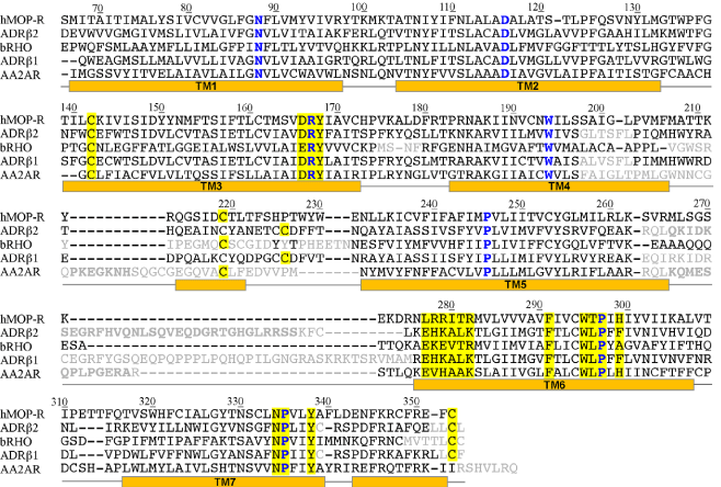
| Figure 1: Sequence alignment used in the creation of the models of the human μ opioid receptor, hMOP-R. The templates are: human β2 adrenergic receptor (ADRβ2), bovine rhodopsin (bRHO), turkey β1 adrenergic receptor (ADRβ1) and human A2A adenosine receptor (AA2AR). The residues of the N- and C- terminal segments are excluded (residues 1 to 65 and residues 354 to 400, respectively). Also, the residues excluded from the comparative modeling are colored in gray. The most conserved residues at each of the transmembrane helices are depicted in blue. The secondary structure of the β2 adrenergic receptor based on STRIDE [32], is shown below the sequences. Residue numbering of hMOP-R is shown. Highly conserved motifs in the rhodopsin-like GPCR family are highlighted in yellow. |