|
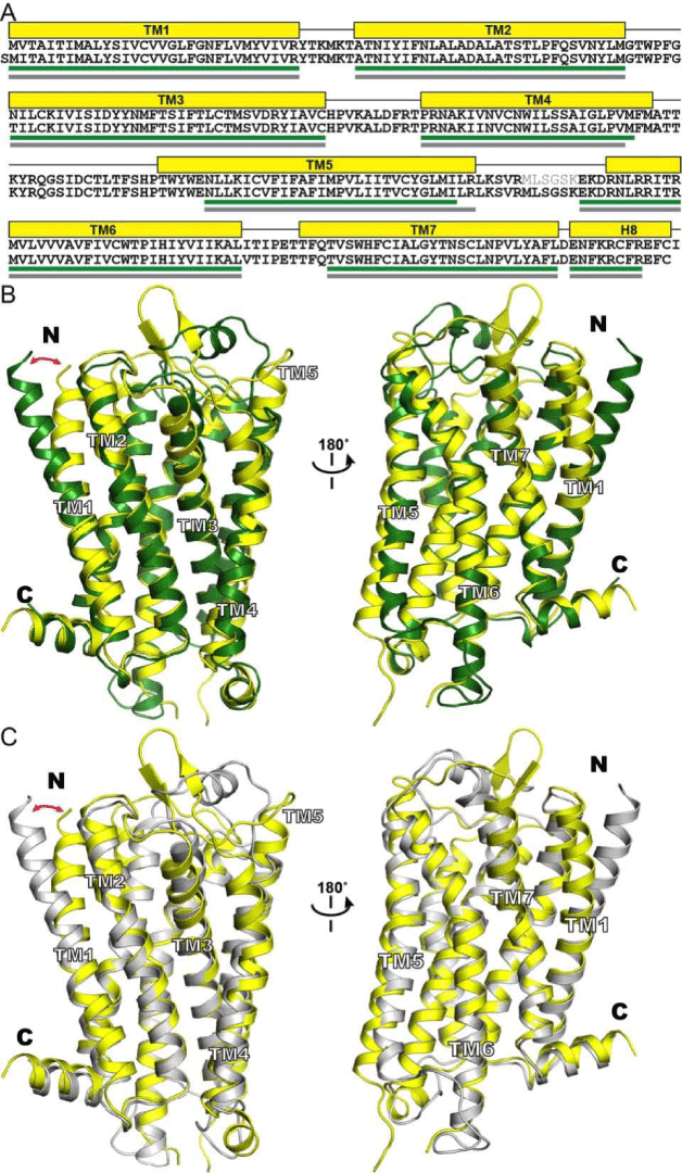
| Figure 3: Comparison of the structure of mouse μ opioid receptor with the models of the human μ opioid receptor 4T-hMOP-R and 2T-hMOP-R. (A) Sequence alignment that displays the length of the TM helices in the crystal structure (top sequence) and the 4T-hMOP-R and 2T-hMOP-R models (bottom sequence). The helical segments are depicted as yellow boxes for the crystal structure and as green and gray lines for 4T-hMOP-R and 2T-hMOPR, respectively. (B) Crystal structure (yellow) and the 4T-hMOP-R model (green) are superimposed. (C) Crystal structure (yellow) and the 2T-hMOP-R model (gray) are superimposed. The red arrow indicates the different position of the extracellular half of the TM1 helix. |