|
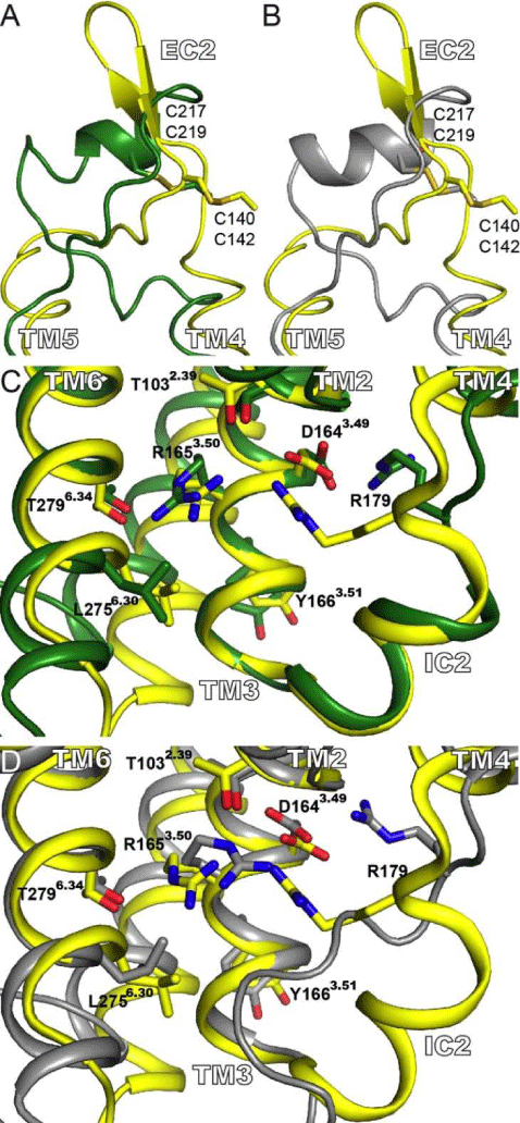
| Figure 4: Structure of the EC2 and IC2. (A) and (B) Superposition of the EC2 for the crystal structure (yellow) and the 4T-hMOP-R (green) and 2T-hMOP-R (gray) models, respectively. The conserved disulfide bond between C140 in TM3 and C217 in EC2 is displayed. The numbers below correspond to the residue number for the equivalent residues in the human μ opioid receptor (C219 and C142). (C) and (D) display views from the intracellular milieu, particularly interactions around the DRY motif. In (C) a comparison of the crystal structure (yellow) with 4T-hMOP-R (green) is shown. In (D) comparison of the crystal structure (yellow) with 2T-hMOP-R (gray) is shown. Relevant side chains are depicted. |