|
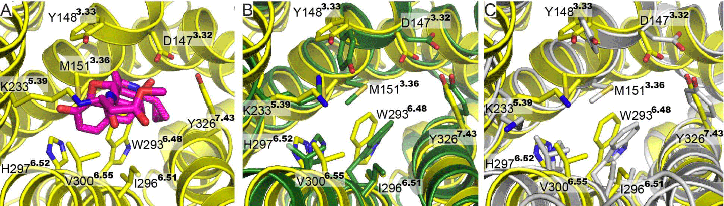
| Figure 5: Binding pocket comparison. (A) The binding pocket of the crystal structure of mouse μ opioid receptor with the structure of β-FNA (magenta) is displayed. The nine residues that directly interact with β-FNA are indicated. (B) and (C) Comparison of the nine residues in the binding pocket of the crystal structure (yellow) and the 4T-hMOP-R (green) and 2T-hMOP-R (gray) models, respectively. |