|
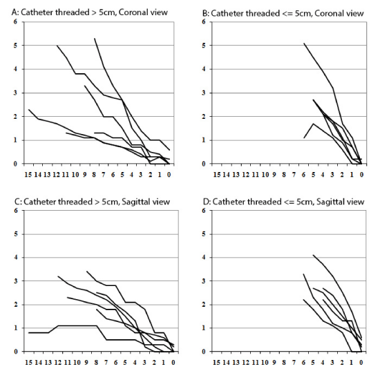
| Figure 2: The Movement of Catheter Tips under Fluoroscope. Each line represents one sample. Sagittal view and coronal view are shown separately. X-axis represents the distance of catheter tip beyond the original Contiplex needle tip as calculated by catheter marks at skin. Y-axis represents the distance of catheter tip to the original Contiplex needle tip as calculated under fluoroscope. |