|
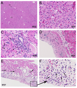
| Figure 3: Histopathology of internal organs (liver and heart) in donors and recipients exposed to Isoflurane alone. Images show histopathology (HE staining) of a donor liver (A, B and C; magnifications 40X, 400X, 400X) and transplanted heart (D, E and F; magnifications 400X, 100X; Inset magnification) in animals that developed malignant hyperthermia. Figures 3A and 3B show congestion of liver sinusoids with greater severity observed in the zone 2 (white arrows) of liver lobules. Higher magnification also revealed the presence of lymphocytic infiltrates (3C; black arrowheads) with occasional eosinophils (3C; white arrowhead) in the region of portal triad. The endocardium of transplanted heart (3D) was devoid of any inflammatory infiltration but the epicardial side revealed infiltrates creeping into the myocardium (3E; black arrows) at several places. Magnified region shows neutrophilic infiltration (3F; black arrows) with occasional eosinophils (3F; white arrowhead). PT: Portal Triad; CV: Central Vein. |