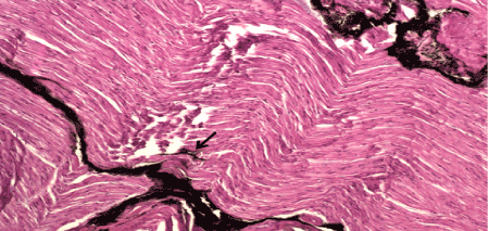
Figure 2:
Histological section of nerve with intra-fascicle injection, showing particulate dye within the fascicle (arrow) as well as disruption of the fascicular elements.