 |
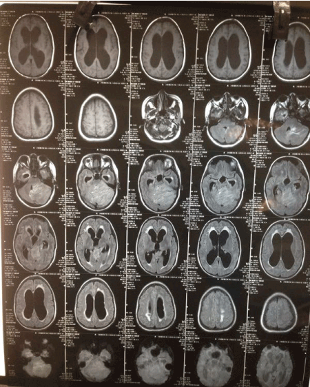
| Figure 1: MR brain imaging reveals a large, ill defined,altered signal intensity, infratentorial mass lesion involving the left cerebellar hemisphere extending upto the region of the vermis with widened cerebellar folli and striated appearance, with apparently preserved cortical striations , with obstructive hydrocephalus, periventricular ooze and B/L optic nerve hydrops. |