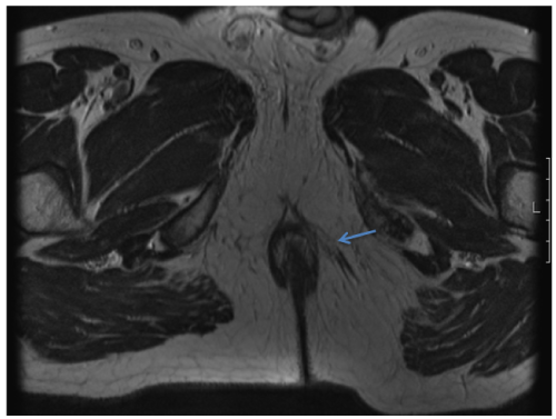
Figure 3:
Diagnostic MRI (supine). Fiber tracts around the left pudendal terminal branches.