|
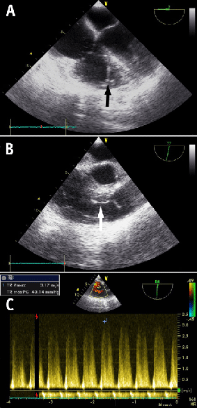
| Figure 2: Still images of video clips and continuous wave Doppler measurement made during the event showing: A. Mid esophageal 4-chamber view showing a dilated right atrium and right ventricle, and a echo dense fragment (black arrow) in the right ventricle. B. Mid esophageal view (70O) showing (white arrow) an approximately 4 cm long and 3 mm wide echo dense fragment in the right ventricular outflow tract. C. Peak measurement of the continuous wave echo-Doppler signal of the tricuspid regurgitant flow of 3m/s, consistent with an elevated peak pulmonary artery pressure of 36mmHg + central venous pressure (Bernoulli equation). |