|
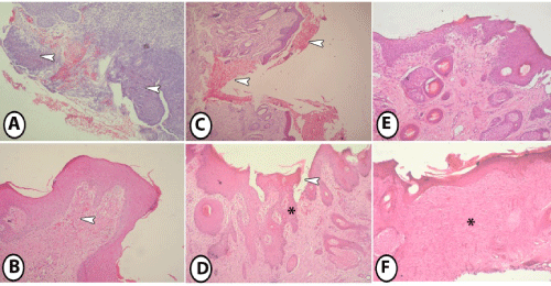
| Figure 3: Histopathological examination. 3A: Control group at 7 days post-wound induction showing infiltration of many inflammatory cells at site of the wound (arrowheads), 3B: Control group at 14 days post-wound induction showing immature granulation tissue (arrowhead), 3C: Lidocaine group at 7 days postwound induction showing extensive hemorrhage (arrowheads), 3D: lidocaine group at 14 days post-wound induction showing incomplete epithelization of epidermis (arrowhead). Note formation of granulation tissue at subepithelial layer (asterisk). 3E: Lidocaine with epinephrine group at 7 days post-wound induction showing partial epithelization of epidermis, 3F: Lidocaine with epinephrine group at 14 days post-wound induction showing presence of mature collagen bundles at the dermis (asterisk). |