 |
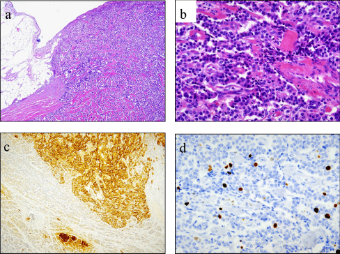
| Figure 2: (a) Histological findings of the pancreatic tumor (hematoxylin-eosin staining, low-power field). (b) Histological findings of tumor cells with anisokaryosis (hematoxylin-eosin staining, high-power field). (c) Immunohistochemistry using antibody against insulin showed positive staining in tumor cells and normal islet cells of the pancreas (insulin stain, low-power field). (d) Immunohistochemical staining for MIB-1 (high-power field). The labeling index was 5%. |