|
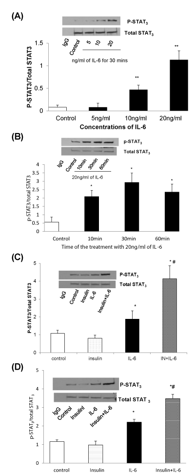
| Figure 2: Chronic insulin treatment enhances IL-6-induced STAT3 tyrosine phosphorylation. Cells were treated with serum-free (Figure 2A and B) medium or serum-free medium containing 100nM (Figure 2C), or 5nM (Figure 2D) of insulin for 24 hours followed by stimulation with IL-6 for indicated concentrations (Figure 2A) or 20ng/ml (Figure 2C and 2D) and indicated times (Figure 2A) or 30min (Figure 2C and 2D), and lysed with RIPA buffer. p-STAT3 was checked by immunoprecipitation with anti-STAT3 and probed with antiphospho- STAT3 (Tyr705). Membranes were stripped and reprobed with anti- STAT3 to serve as a loading control. Upper panels show the representative Western blot. Graphs show mean±SE of the ratio of phosphorylated STAT3 to total STAT3 from three independent experiments. Data were analyzed by using One-Way ANOVA followed by post hoc test, *P<0.05, **P<0.01, compared to control and insulin. #P<0.05 compared to IL-6 alone. |