|
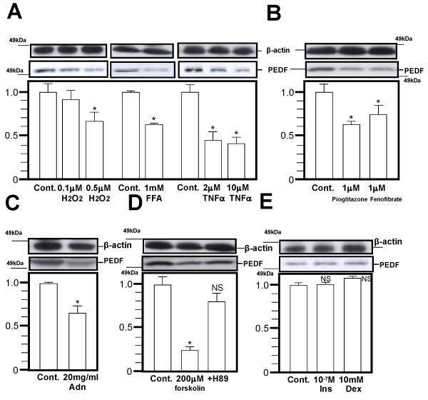
| Figure 2: PEDF expression (western blot; upper panel, Q-PCR; lower panel) in 3T3-L1 adipocytes with 12 hr incubation using H2O2, FFA and TNFα (A), PPAR agonists (B), adiponectin (C), PKA agonists (D), insulin and dexamethasone (E). The mature 3T3-L1 adipocytes (days 6-10) were incubated with the indicated chemicals for 12 hr, followed by western blot (upper panel) and Q-PCR (lower panel). Representative data from four independent experiments are presented. *Significant difference (P <0.05) relative to PEDF expression in control cells. N.S.; not significant relative to control cells. |