|
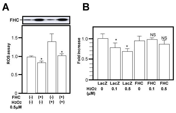
| Figure 4: PEDF expression with FHC overexpression. The adenovirus expressing Lac Z or FHC was transfected into 3T3-L1 adipocytes and levels of FHC protein expression were confirmed (A, upper panel). ROS levels in the presence or in the absence of 0.5 μM H2O2 were measured flow cytometrically by determining the mean fluorescent intensity relative to that of the control group (A, lower panel). PEDF expression in the cells was examined by Q-PCR (B). Representative data from four independent experiments are presented. *Significant difference (P <0.05) relative to control cells. N.S.; not significant relative to control cells. |