 |
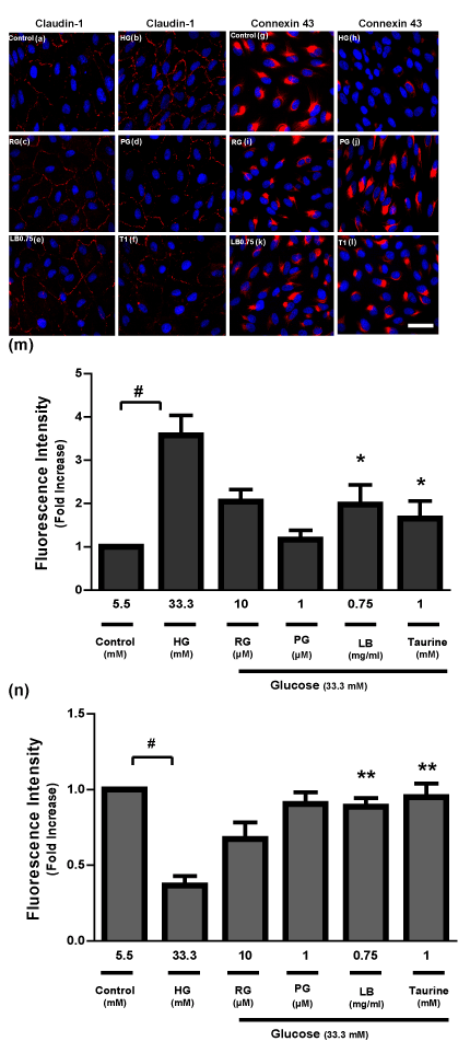
| Figure 8 : Immunohistochemistry of ARPE-19 cells showing the distruption of the monolayer induced by high glucose and the effects of LB extract (0.75 mg/ml) and taurine (1 mM) in preventing the disorganization of claudin-1 and connexin 43 proteins and in maintaining the integrity of the monolayer. (a-f) claudin-1 and (g-l) connexin 43 staining appears in red. The intensity of (m) claudin-1 and (n) connexin 43 staining signals (red) was quantified using the Image J software. Levels in control were arbitrarily assigned a value of 1.0. Values are means ± SEM (n=4). #P<0.01 versus Control, *P<0.05, **P<0.01 versus high glucose (HG). The nuclei were stained with Hoechst 33342 (blue). Scale bar 50 ìm. HG, high glucose (33.3 mM); RG, RG (10 ìM); PG, PG (1 ìM); T1, taurine 1 mM; LB0.75, LB (0.75 mg/ml). |