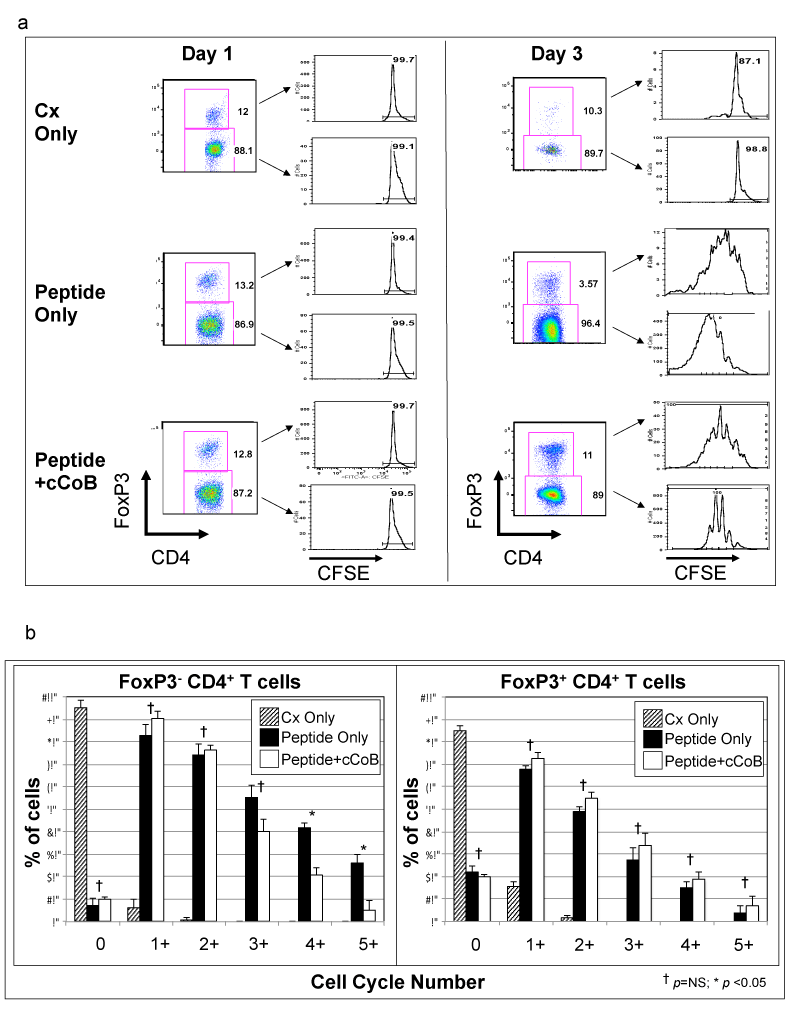
(A) At 1 and 3 days cells were analyzed for CFSE content to determine cell divisions. At the indicated days cells cultured as indicated were stained with anti-CD3, CD4, and FoxP3 and assessed by flow cytometry. CD3+, CD4+ cells were separated based on FoxP3 positivity and CFSE content was assessed in the indicated T cell subpopulations. Shown are representative histograms. (B) Cell cycling was determined by quantitating the percentage of cells that had processes though the indicated generations (i.e. 0 = no divisions, 1+ = at least 1 cell division, etc). Means and standard deviations (error bars) of percentages of cell subpopulations from 3 experiments are shown. ANOVA analysis was conducted on the 3 treatment groups (culture only, peptide only, peptide+cCoB) in each generation grouping. Post hoc Student’s t-Test was used to compare percentage of cells in the peptide only and peptide+cCoB groups and p values of >0.05 (NS) or <0.05 are indicated using † and *, respectively.