|
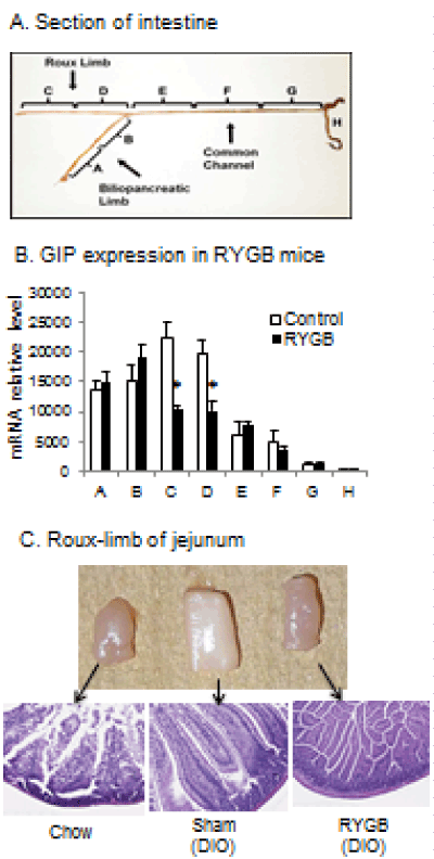
| Figure 4: GIP mRNA in DIO mice after RYGB surgery (Cohort 2). GIP mRNA was measured in mice at 6 wks after surgery when the body weight was stabilized. The intestine samples were collected in the non-fasting condition. A. Intestine fragments are indicated in the diagram. B. GIP mRNA in different intestinal sections after RYGB surgery. Data is presented as mean ± SE (n=5). *p< 0.05 RYGB vs. sham by student’s t test. C. Histology of small intestine. The intestine fragments corresponding to Roux-limb are shown with microscope images (10X) of HE staining. |