|
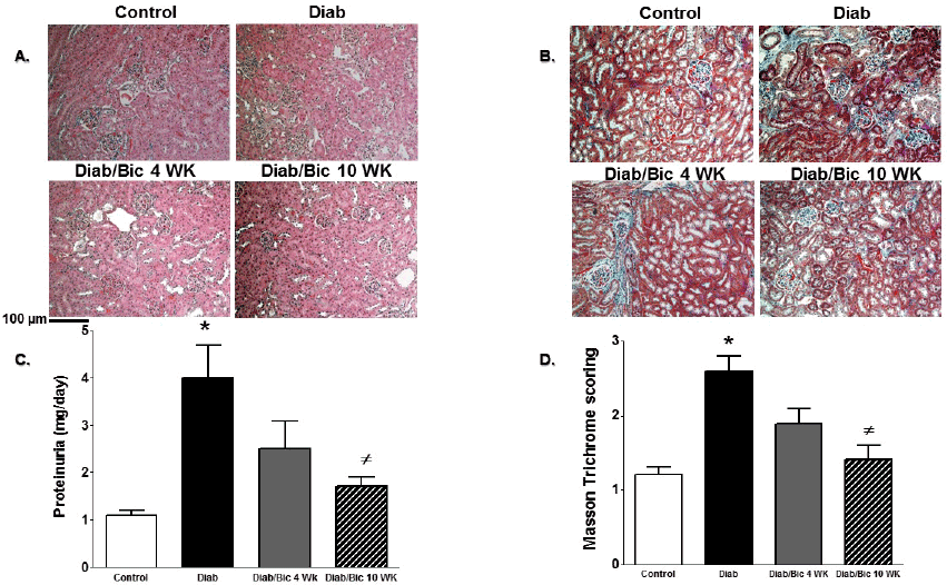
| Figure 4: Representative images (200X) of kidney sections H & E staining (A) and Masson’s trichrome staining (blue staining, B), proteinuria (C) and average score for Masson’s trichrome staining (D) in control and diabetic mice with or without baicalein treatment for 4 and 10 weeks. (*P<0.05 vs. control mice and #P<0.05 vs. diabetic mice; n=4/group for histology and n=6- 8/group for proteinuria). |