|
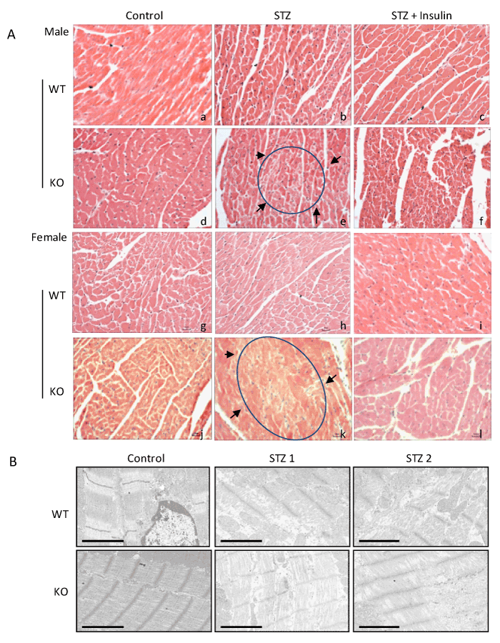
| Figure 2: Myocardial structural damage. Hearts from Nrf2 WT and Nrf2 KO mice were collected 14 days after STZ treatment. Diabetic condition was confirmed by blood glucose detection (≥250 mg/dL). (A) H&E staining: Paraffin-embedded heart tissue (transverse section) was stained with H&E and examined under light microscope (40X). Apparently damaged areas were located inside of circles and arrows. Bar = 20 μm. (B) Ultrastructural damage: Micrographs of transmission electron microscopy of control and diabetic heart samples. Bar = 2 μm. |