|
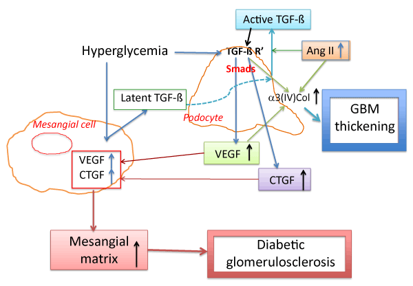
| Figure 2: Schematic illustration of proposed mechanism of TGF-β action inpodocytes and mesangial cells leading to the development of diabetic nephropathy. High glucose stimulates TGF-β secretion in mesangial cells and TGF-β receptor (TGF-β R’) expression in podocytes. Soluble forms of latent TGF-β complex released from mesangial matrix may be localized to the podocyte surface and activated by angiotensin (Ang) II. Activated TGF-β/ Smad signaling pathway in podocytes mayinduce α3(IV) collagen, connective tissue growth factor (CTGF) and vascular endothelial growth factor (VEGF) overexpression, leading to glomerular basement membrane (GBM)thickening and mesangial matrix expansion, culminating in diabetic glomerulosclerosis. |