|
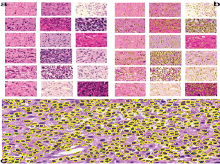
| Figure 2: Cell segmentation result (a) orginal gliomas slides (b) segmention with boundaries of the cell nuclei (c) detail of one segmentation(marked as yellow); Figure 2a shows 16 out of 28 orginal gliomas slides; Figure 2b shows the 18 segmentation results with boundaries of the cell nuclei; Figure 2c shows a detail of one particular segmentation result. |