|
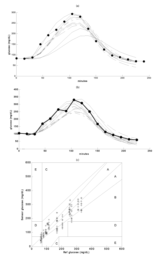
| Figure 7: Multi-day (168-hour) swine experiment. The top panel (7a and 7b) shows day1 and 7 glucose excursion respectively, with 9 sensors (21-gauge, 400-micron fiber optic sensor with PEGDMA: MAA matrix and acrylodanlabeled GGBP) placed subcutaneously (>3mm). The bold line with solid circles displays reference blood glucose as measured with a YSI clinical analyzer. The bottom panel (7c) shows day 7data plotted on a Clarke-error grid. |