|
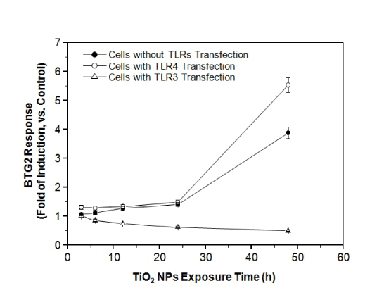
| Figure 2: Time course of HepG2 exposure to TiO2 NPs. The cells were transfected with BTG2 promoter-reporter plasmid and co-transfected with or without TLR3 and TLR4 expression vectors. The transfected cells were exposed to 10 μg/mL TiO2 NPs for the indicated lengths of time. Each plot was produced from at least 3 replicate measurements. All values are presented as mean ± S.D. (n ≥ 3). |