|
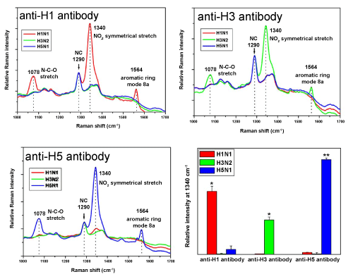
| Figure 4: SERS spectra of the half sandwich assay with three different kinds of influenza H1N1-VLPs (red), H3N2-VLPs (green), and H5N1-VLPs (blue) against a series of antibodies. The Raman signal at 1290 cm-1 from the NC membrane is consistent and used as an internal standard here. Bar graphs show the intensities of the SERS peaks at 1340 cm-1 for different subtypes of influenza VLP. |