|
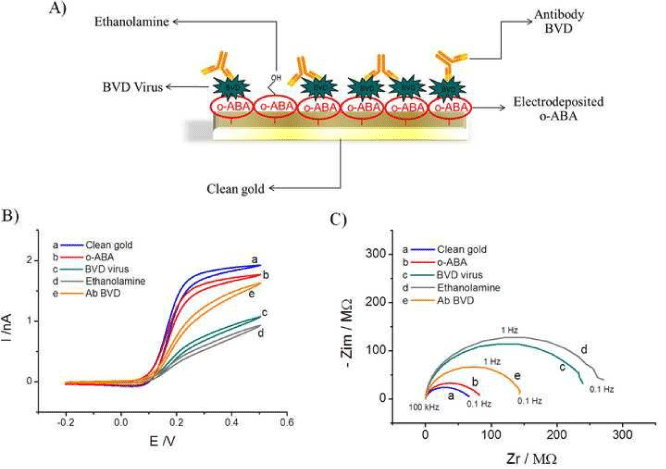
| Figure 4: (A) Schematic of the layer-by-layer build up on a nanowire sensor for BVD antibody detection; (B) Cyclic Voltammograms and (C) Nyquist plots of (a) pristine gold nanowire electrode; (b) o-ABA modified electrode; (c) BVD virus (100 μg/mL) modified electrode; (d) ethanolamine blocked electrode; (e)Antibody BVD (10 μg/mL) binding in buffer. Solution composition: 1 mMFcCOOH in 10 mM PBS. CV scan rate: 100 mVs−1. EIS frequency range: 0.1 Hz to 100 kHz; E=150 mV and ΔV=5 mV. |