|
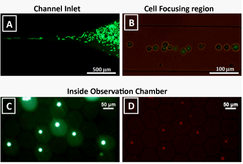
| Figure 3: (A) Live cells entering the microchannel stained with calcein- AM dye; (B) Cells focused to the centre of the microchannel due to the inertial effects. Droplets collected in the observation chamber viewed with representative (C) green and (D) red emission filter cubes to show live cells whose membrane was just lysed thus allowing EthD-1 to enter inside them. |