|
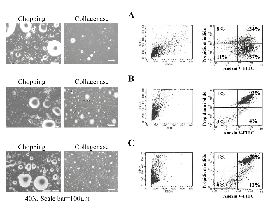
| Figure 2: Analysis of cell death in cells isolated from preserved adipose tissues without cryopreservation solution. Human adipose preserved in −20°C without cryopreservation solution for 1, 3, or 12 months. Cells from adipose tissues isolated by collagenase were cultured in media (DMEM containing 10% FBS). Analysis of annexin V and PI staining of cells isolated from human adipose tissues preserved at −20°C without cryopreservation solution for 1, 3, or 12 months. Annexin V and PI staining was analyzed by flow cytometry. Surviving cells, for which both annexin V and PI levels are low, are shown in the lower left quadrant. Data from a representative experiment (from a total of three) are shown. A) Left: Image of human adipose cells and Right: analysis of annexin V and PI staining cells isolated from human adipose tissues preserved at −20°C without cryopreservation solution for about 1months. B) Left: Image of human adipose cells and Right: analysis of annexin V and PI staining cells isolated from human adipose tissues preserved at −20°C without cryopreservation solution for about 3months. C) Left: Image of human adipose cells and Right: analysis of annexin V and PI staining cells isolated from human adipose tissues preserved at −20°C without cryopreservation solution for about 12 months. Scale bar = 40×, 100 μm. |