|
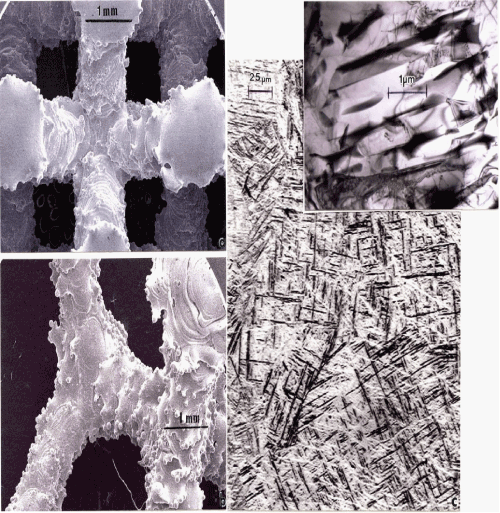
| Figure 8: SEM views for dode-thin mesh strut structure (a) and foam ligament structure (b) corresponding to open-cellular components shown in figure 5c. (c) Shows a light-optical image for α’-martensite structure in the mesh struts in (a). The insert in (c) shows a corresponding TEM image. |