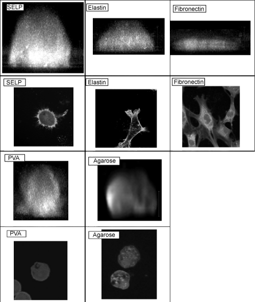
Figure 7:
Fluorescent staining images of actin cytoskeleton of cells cultured for 24 hr on dishes coated with different substances. The coating concentration is 10 μg/ ml. White bar, 10 μm.