|
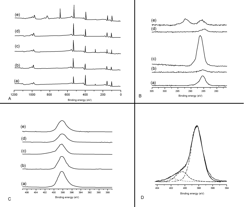
| Figure 2: XPS analysis of MEAs before and after surface modification. A: XPS measurements of the MEA as received (a), the MEA after O2 plasma cleaning treatment (b), with DETA (c) and 13 F (e) treatments. As a control, the MEA surface was also examined after exposing it to LASER irradiation (d). 1s, Si 2p, N 1s and O 1s high resolution spectra for the same set of samples (a-e) were also recorded and presented in B and C. |